Back Bones Diagram : Patient Education Spine Diagrams New York Back Doctor : Bone structure of the femoral head 12 photos of the bone structure of the femoral head bone structure cross sectional view of the femoral head, bone, bone structure cross sectional view of the femoral head
Back Bones Diagram : Patient Education Spine Diagrams New York Back Doctor : Bone structure of the femoral head 12 photos of the bone structure of the femoral head bone structure cross sectional view of the femoral head, bone, bone structure cross sectional view of the femoral head. Strong muscles and bones, flexible tendons and ligaments, and sensitive nerves contribute to a healthy spine. At the back of the vertebral body are bony arches that project outward to form the facet joints and spinous processes. At the back of each bone in the spine (vertebra) are bony points called processes, which muscles attach to. The first seven bones (vertebrae) of your spine form your neck. These problems can reduce the amount of space available for your spinal cord and the nerves that branch off it.
The vertebral column houses the spinal canal, a cavity that. The bones of the pelvis and lower back work together to support the body's weight, anchor the abdominal and hip muscles, and protect the delicate vital organs of the vertebral and abdominopelvic cavities. The temporal bones are located lateral to the temporal lobes of the brain, and each bone consists of five parts. Bone structure of the femoral head 12 photos of the bone structure of the femoral head bone structure cross sectional view of the femoral head, bone, bone structure cross sectional view of the femoral head Your medication, delivered learn more > customers also shopped for.
For more anatomy content please follow us and visit our website:
We are pleased to provide you with the picture named anatomy of back muscles diagram.we hope this picture anatomy of back muscles diagram can help you study and research. This article looks at the anatomy of the back, including bones, muscles, and nerves. Lateral labeled diagram of the human vertebral spinal column showing vertebrae numbering order and the 5 different regions of the spine. These bones are connected at the back with specialized joints. Individual anatomical structures include 2: A tough, springy disc of cartilage sits between the vertebrae of your spine. As your spine ages, it's more likely to experience bone spurs or herniated disks. The vertebrae, which stack like spools of thread, support the back and protect the spinal cord. Can you feel the bumps of your vertebrae along your back? The temporal bones are located lateral to the temporal lobes of the brain, and each bone consists of five parts. The lower part of the trapezius ascends and depresses the scapula, while the transverse or middle region of the trapezius is what retracts the. All the images are in vector format, allowing an optimal web display with zoom and shifting of the anatomical images. People also love these ideas.
The lower part of the trapezius ascends and depresses the scapula, while the transverse or middle region of the trapezius is what retracts the. At the back of each bone in the spine (vertebra) are bony points called processes, which muscles attach to. Diagram hand bones anatomy 12 photos of the diagram hand bones anatomy , bone. Individual anatomical structures include 2: Related posts of human back bones diagram bone structure of the femoral head.

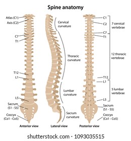
The spine diagram the spine diagram shown below, consists of many bones or vertebrae,soft discs,the spinal cord, and spinal nerves.
Your medication, delivered learn more > customers also shopped for. At the same time the bones grow larger by growing back into the growth plates. The spine anatomy is a complex structure. This process continues until the end of puberty, when the growth plate stops growing and the bones fuse permanently into a single bone. These bones work together to provide. The red lines point individual bones and the names are writen in singular, the blue lines conect to group of bones and are in plural form. Spinal anatomy is a remarkable combination of strong bones, flexible ligaments and tendons, large muscles and highly sensitive nerves. Diagram of a human female skeleton, back view. These bones are connected at the back with specialized joints. People also love these ideas. Tumors that begin in the bones of the spine (primary tumors) are far less common. A tough, springy disc of cartilage sits between the vertebrae of your spine. Vertebrae separated by intervertebral discs.
The atlas is the topmost vertebra, and along with c2, forms the joint connecting the skull and spine. Related posts of human back bone chart diagram hand bones anatomy. There are three parts to the trapezius. The spinal cord begins at the base of the brain and extends into the pelvis. The vertebral column, also known as the backbone or spine, is part of the axial skeleton.the vertebral column is the defining characteristic of a vertebrate in which the notochord (a flexible rod of uniform composition) found in all chordates has been replaced by a segmented series of bone:

There are three parts to the trapezius.
There are three parts to the trapezius. This is a single bone that is present at the back and lower part of the cranium, just behind the parietal and temporal bones. Related posts of human back bones diagram bone structure of the femoral head. Page 1 of 1 start over page 1 of 1. Diagram of a human female skeleton, back view. The trapezius or trapezoid muscles are two paired muscles that extend from the base of the thoracic vertebrae in the spine to the occipital bone and run out to the spine of the scapula. The vertebral column of the lower back includes the five lumbar vertebrae, the sacrum, and the coccyx. We are pleased to provide you with the picture named anatomy of back muscles diagram.we hope this picture anatomy of back muscles diagram can help you study and research. The lower part of the trapezius ascends and depresses the scapula, while the transverse or middle region of the trapezius is what retracts the. This is a tutorial to quickly s. Multiple myeloma is a type of cancer that often metastasizes to the spine. Vertebrae separated by intervertebral discs. The vertebral column houses the spinal canal, a cavity that.


Komentar
Posting Komentar